Business Wire01.19.18
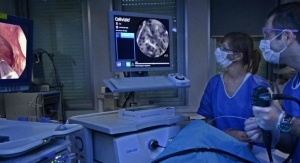
Mauna Kea Technologies, inventor of Cellvizio, the multidisciplinary confocal laser endomicroscopy (CLE) platform, has received its 14th U.S. Food and Drug Administration (FDA) 510(k) clearance for the Cellvizio 100 series and all of its Confocal Miniprobes.
THe FDA agreed that data from multiple peer-reviewed medical journals validate the technology’s ability to image the internal microstructure of tissues including, but not limited to, the identification of cells and vessels and their organization or architecture. The addition of the indications for: “identification of cells and vessels and their organization or architecture” to the previously cleared intended use (the fields of gastroenterology,1 urology,2 and pulmonology3 during endoscopic, laparoscopic manual and robot-assisted surgical, and image-guided percutaneous procedures), represents a pivotal milestone for in vivo real-time microscopic visualization technology.
“With this new clearance, Cellvizio is affirmed as the only platform for imaging and identification at the cellular and micro-vascular level during a wide variety of procedures in endoscopy and surgery,” said Sacha Loiseau, Ph.D., founder and CEO of Mauna Kea Technologies. “This milestone enables us to formally shift from imaging to identification, providing us with strong leverage to broaden adoption by healthcare providers and payers.”
This new clearance builds on the recent executive summary published by the College of American Pathologists (CAP) which highlighted the potential value of in vivo microscopy (IVM). The summary noted that “the architectural and cellular patterns generated by IVM are interpretable by pathologists to make differential diagnoses and to identify areas for biopsy, improving diagnostic yield. Such directed biopsies decrease sampling errors resulting in fewer, less frequent biopsies, significantly decreased morbidity, and significant cost saving […]”4
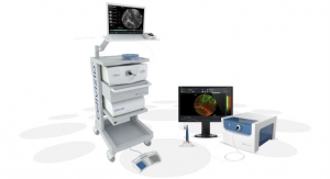
Mauna Kea Technologies is a global medical device company focused on eliminating uncertainties related to the diagnosis and treatment of cancer and other diseases thanks to real time in vivo microscopic visualization. The company’s flagship product, Cellvizio, has received clearance/approval in a wide range of applications in more than 40 countries, including the United States, Europe, Japan, China, Canada, Brazil, and Mexico.
References
1. Wallace, M., Lauwers, G., Chen, Y., Dekker, E., Fockens, P., Sharma, P., & Meining, A. (2011). Miami classification for probe-based confocal laser endomicroscopy. Endoscopy, 43(10), 882–891. https://doi.org/10.1055/s-0030-1256632
2. Chen, S. P., & Liao, J. C. (2014). Confocal Laser Endomicroscopy of Bladder and Upper Tract Urothelial Carcinoma: A New Era of Optical Diagnosis? Current Urology Reports, 15(9). https://doi.org/10.1007/s11934-014-0437-y
3. Thiberville, L., Salaun, M., Lachkar, S., Dominique, S., Moreno-Swirc, S., Vever-Bizet, C., & Bourg-Heckly, G. (2009). Human in vivo fluorescence microimaging of the alveolar ducts and sacs during bronchoscopy. European Respiratory Journal, 33(5), 974–985. https://doi.org/10.1183/09031936.00083708
4. http://www.cap.org/ShowProperty?nodePath=/UCMCon/Contribution%20Folders/WebContent/pdf/in-vivo-executive-summary.pdf
THe FDA agreed that data from multiple peer-reviewed medical journals validate the technology’s ability to image the internal microstructure of tissues including, but not limited to, the identification of cells and vessels and their organization or architecture. The addition of the indications for: “identification of cells and vessels and their organization or architecture” to the previously cleared intended use (the fields of gastroenterology,1 urology,2 and pulmonology3 during endoscopic, laparoscopic manual and robot-assisted surgical, and image-guided percutaneous procedures), represents a pivotal milestone for in vivo real-time microscopic visualization technology.
“With this new clearance, Cellvizio is affirmed as the only platform for imaging and identification at the cellular and micro-vascular level during a wide variety of procedures in endoscopy and surgery,” said Sacha Loiseau, Ph.D., founder and CEO of Mauna Kea Technologies. “This milestone enables us to formally shift from imaging to identification, providing us with strong leverage to broaden adoption by healthcare providers and payers.”
This new clearance builds on the recent executive summary published by the College of American Pathologists (CAP) which highlighted the potential value of in vivo microscopy (IVM). The summary noted that “the architectural and cellular patterns generated by IVM are interpretable by pathologists to make differential diagnoses and to identify areas for biopsy, improving diagnostic yield. Such directed biopsies decrease sampling errors resulting in fewer, less frequent biopsies, significantly decreased morbidity, and significant cost saving […]”4
Mauna Kea Technologies is a global medical device company focused on eliminating uncertainties related to the diagnosis and treatment of cancer and other diseases thanks to real time in vivo microscopic visualization. The company’s flagship product, Cellvizio, has received clearance/approval in a wide range of applications in more than 40 countries, including the United States, Europe, Japan, China, Canada, Brazil, and Mexico.
References
1. Wallace, M., Lauwers, G., Chen, Y., Dekker, E., Fockens, P., Sharma, P., & Meining, A. (2011). Miami classification for probe-based confocal laser endomicroscopy. Endoscopy, 43(10), 882–891. https://doi.org/10.1055/s-0030-1256632
2. Chen, S. P., & Liao, J. C. (2014). Confocal Laser Endomicroscopy of Bladder and Upper Tract Urothelial Carcinoma: A New Era of Optical Diagnosis? Current Urology Reports, 15(9). https://doi.org/10.1007/s11934-014-0437-y
3. Thiberville, L., Salaun, M., Lachkar, S., Dominique, S., Moreno-Swirc, S., Vever-Bizet, C., & Bourg-Heckly, G. (2009). Human in vivo fluorescence microimaging of the alveolar ducts and sacs during bronchoscopy. European Respiratory Journal, 33(5), 974–985. https://doi.org/10.1183/09031936.00083708
4. http://www.cap.org/ShowProperty?nodePath=/UCMCon/Contribution%20Folders/WebContent/pdf/in-vivo-executive-summary.pdf